Transcription
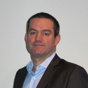
Olivier Delporte 0:05
Thank you. Okay, thank you for the organizers. Good morning, everyone. So, we're going to talk about SamanTree Medical that I'm the CEO of since a couple of months now. Samantree'sabout cancer, cancer surgery, and specifically, tissue assessments in the operating room tissue assessment. So we were in interventional radiology a few minutes ago, we're going to stay within the cancer space. We're going to talk about what SamanTree does, we're doing fresh tissue assessments. So basically, the tumor excise specimen to be analyzed in real time in the operating room in a variety of cancer surgeries. And so what that allows to and what really the goal is to guide treatment plan during surgery and reduce time. How do we do that? So the company has developed the histologic scanner, which is based on confocal microscopy, confocal microscopy, which exists for decades, but has been evolved to basically allow for a ultra fast in the operating room in real time, high resolution image. So in 45 to 50 seconds. What the scanner provides is a histology like picture of the surface of a specimen with no cutting, no staining, which means the specimen remains open for any subsequent pathology or pathology related assessment. So it's margin assessments in the operating room in real time. A number of you have probably heard about margin assessment or intraoperative margin assessment, which is some referred to has been a holy grail of, you know, cancer surgeries. Well, this is really in real time margin assessment. If we think about what does it replace, or how does it fit into the workspace, the workflow? Well, until today, basically when there is a cancer surgery, and there is tumor excision. Well, in the operating room, either the surgeons are blind, and they will know a week later or two weeks later from histology, if there were what's called positive margins, in which case, the patient will need either to be reintervened on in will eventually or with chemotherapy, for example, or the pathologist can also do what's called frozen section analysis, which is complex wise resources wise, it takes about 40 minutes involves pathologist and sometimes for logistics purposes, the pathology lab is not even in the same building. So there are hospitals where it takes more than an hour. So it's slow resources demanding. And what the Histalog allows is basically, during the surgery in the operating room in five minutes, to have a histology like image for a surgeon or a pathologist to interpret and provide guidance to the surgeon. IE did the surgeon cut, not too much, but enough? Or should the surgeon cut a little more? So what is really the product about besides the scanner? Well, there is a scanner, which is a piece of capital equipment. It's imaging as you've seen, we have consumables a dish and a dip that are proprietary, but also a digital solution. And the digital solution allows for one Remote Connectivity. So a pathologist or an image reviewer can take control of the console, which is in the operating room from a distance. So that means workflow efficiencies for pathologist. We are also working on AI, as artificial intelligence will help will enhance the human eye. And so in breast cancer specifically, we have an AI software coming up in a couple of months. And finally, we're working on DICOM connectivity or PACs integration for archiving purposes to have a complete digital suite integrate. So, looked at it differently. We're talking about interventional oncology, if you look at what tools are available in the EU are like ultrasound ultrasound guided surgery. What the Histolock image assessment technology allows is to have an histology like image, high resolution, we're talking of might one to two microns in real time in the operating room. What if you know what if we used the Histolock and what is it used for which cancer surgeries? Well, the the the one of the key advantages is that it can be used in a lot of cancer surgeries. The large applications or use cases are breast cancer, prostate cancer just by the patient number and the unmet need. Yet, the Histolock has been used for kidney cancer, lung cancer, head and neck cancer, skin cancers and brain. And again, it's always about in the operating room assessing if the tissue is cancerous and if there are cancerous cells on a specimen or even on a biopsy, so on a small elements competitive landscape. So this is a this slide shows basically, range of specialties, which means in which use cases can it be used, versus time, and we're talking of a few minutes for a variety of use cases, which, you know, makes us believe that we are very differentiated from a number of other companies active in the space, allowing for all the use cases that I showed to you from prostate and breast cancer to lung cancer, brain cancer. And it's really thanks to the combination of the speed, the resolution and the accuracy of the images. A few words about the unmet needs. So the unmet need in breast is clear. Today, 25 to 30% of women undergoing breast cancer, conservative surgery have positive margins, meaning they're going to be re intervened on. Our device has been shown and it's published that use then eventually it can reuse the RE intervention rate by 75%. So that 25 to 30% would be reduced by 75%. In prostate, it's about keeping continents sexual function, which are today issues in 20 to 40% of the robot assisted radical prostatectomies do the histologic has been shown to be accuracy why similar to frozen section analysis yet in 80%, less time, save 30% or 30 minutes of an operating room time, there is a significant saving there in value proposition. So that's the unmet need, which leads to large markets. We're talking of multibillion dollar markets between the healthcare system savings on the pathology side and the improvement in clinical outcomes in a variety of use cases. So large markets. Finally, the device has been used in as of today, close to 2500 patients in a variety of use cases, as you see breast, prostate skin, others published in nine publications today, and systematically showing high performance or high accuracy. Fast. So in less than 60 seconds the image is acquired and easy training and easy workflow. And not only the pathologist can learn to review the images but the surgeons can so today we have in some hospitals, surgeons performing breast surgeries who make a decision of reinventing ie cutting further or not based on the image and they don't need only either radiologist or pathologist business model and sales traction. So the business model is typical for capital equipment on one hand, so sold or rented console, we have the consumables that are proprietary and sold per surgery, there is software with the Remote Connectivity and AI that will be sold as a license as of next year and service and maintenance. So typical workstreams for a device like that the company started selling last year, and is enjoying very positive sales traction. Happy to talk about that and numbers offline. And if we put it all into a Strat map, the future is about growing sales in Europe, getting access to the US and launching in the second part of next year. Continuing clinical studies, there are a number of interventional studies, interventional studies going on. In breast in prostate we have new data presented last week in brain and in lung and continue those further as well as launch the digital Suite products between now and next year. And finally, because my time is up, the team is very diverse, very experienced in the imaging space very fortunate to have the team and finally we are here. Because we are raising funds. We're going to raise 10 to 15 plus million Swiss francs or euros to support commercialization get 510K clearance in the US prepare for a US go to market strategy, some clinical studies and finalize or will finish product development and r&d. Thank you very much, happy to talk offline.
Market Intelligence

Schedule an exploratory call
Request Info17011 Beach Blvd, Suite 500 Huntington Beach, CA 92647
714-847-3540© 2024 Life Science Intelligence, Inc., All Rights Reserved. | Privacy Policy
